Embryology of the chicken
Embryology of the chicken
The embryology of the chicken is the development of the chicken inside of the egg. Hatchery managers need to be able to differentiate between normal and abnormal embryos and identify possible causes of embryo mortality during incubation.
Fertilisation and early development
Three stages of the early development of the chicken embryo
Fertilisation of the germinal disc by the sperm takes place in the infundibulum about 15 minutes after its holding follicle has released the yolk. Cell division to create the new embryo starts about five hours after fertilisation and continues while the egg passes along the oviduct and after the egg is laid. It is generally said that the hen’s egg takes 21 days of favourable incubation conditions for the chicken to develop and hatch. However, this development takes 22 days – one day in the oviduct and 21 days in the incubator or nest.
The zygote
When the sperm cell (with half the required chromosomes) fertilises the female egg cell (with the other half of the required chromosomes) it forms the zygote, which is a single cell with the correct number of chromosomes. About five hours after fertilisation the zygote enters the isthmus and it is here that the new embryo starts to develop by simple cell division. By the time the egg leaves the isthmus, the zygote, now called the blastoderm or embryo, comprises eight cells and after four hours in the uterus it has grown to 256 cells.
Formation of ectoderm, endoderm and mesoderm
Initially, the dividing cells form one layer over the yolk, but as cell division continues two layers are formed. These are called the ectoderm (uppermost) and the endoderm (underneath) layers. At about this stage the central cells of the blastoderm separate from their contact with the yolk to form a cavity. It is in this cavity that subsequent embryo development occurs. Soon after the formation of the ectoderm and endoderm, a third layer of cells called the mesoderm, or middle layer, is formed.
Early development of the chick embryo (2 days)
From this stage on, the organs and tissues of the bird will develop from these three layers of cells.
- The ectoderm produces the nervous system, parts of the eyes, the feathers, beak, claws and skin.
- The endoderm produces the respiratory system, the digestive system and secretory organs.
- The mesoderm produces the skeleton, muscles, circulatory system, reproductive organs and excretory system.
Another important development at this stage is the way the cells change to allow the production of the different types of cells that make up the tissues. By the time the egg is laid the embryo consists of many cells differentiating into the various tissues, organs and body systems.
Physiological zero
The fowl retains some vestiges of the characteristics of its reptilian ancestors. One characteristic in particular is the influence of ambient temperature during the post laying period on embryonic development. When the temperature of the egg is below 20°C, the embryo becomes dormant and most development stops. When the temperature rises above 20°C, embryonic activity re-initiates. This temperature of about 20°C when embryonic activity starts or stops is often referred to as a physiological zero.
Fluctuating temperatures above/below 20°C will create a start/stop response in embryonic development, and each succeeding response progressively weakens the embryo. The temperature must be increased to the required 37-38°C for optimum development to occur. Failure to satisfy this need leads to significantly weaker embryos. To retain maximum viability of the embryo, hatching eggs should be processed and placed in cool storage below 20°C as soon as possible after collection and held at that temperature until the pre-warming process just prior to setting the eggs in the incubator. Once in the incubator, the temperature must be controlled within very close parameters.
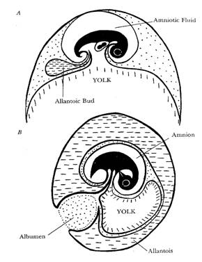
Extra embryonic membranes of the chicken
Extra-embryonic membranes
Because the avian embryo has no anatomical connection to the hen, all of its nutritive requirements, except oxygen, must be contained within the egg. From very early on, the embryo develops special membranes external to its body to access the nutrients in the egg and to carry out essential bodily functions.
There are four of these special membranes and their names and functions are as follows:
- Yolk sac: This sac envelops the yolk and produces an enzyme that changes the yolk material into a form that can be used as a food source by the developing embryo. Any remaining, unused yolk material in the yolk sac when the chicken hatches from the egg is drawn into the abdomen for use by the chicken for the first two to three days after hatching while the chicken learns what to eat/drink and where to find it.
- Amnion: The amnion forms a sac that is filled with fluid in which the embryo floats. In this way it provides a shock-absorbing environment in which the fragile embryo can develop without harm from normal day to day knocks.
- Allantois: The allantois develops an extensive circulatory system connected to that of the embryo and is driven by the new embryonic heart. When the allantois is fully developed it completely surrounds the embryo. This membrane has a number of functions:
- Respiratory – the developing embryo uses oxygen and produces carbon dioxide (it respires). It is unable to carry out this function for itself and hence the allantois oxygenates the blood and eliminates carbon dioxide.
- Excretory – it removes the wastes that result from the embryo’s metabolism and deposits it in the allantoic cavity.
- Digestive – it provides the means for the embryo to access the albumen and the calcium of the shell.
- Chorion: The chorion fuses the inner shell membrane to the allantois and helps that membrane to carry out its functions.
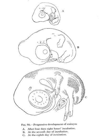
Daily embryonic development
To better carry out an investigation into poor hatchability it is necessary to have knowledge of the way the embryo develops from day to day. This allows the hatchery manager to determine at what age/stage embryos may have died. This is important information when attempting to identify the cause of any poor results.
Photographs of the following steps may be viewed by clicking here.
- DAY 1: Appearance of embryonic tissue.
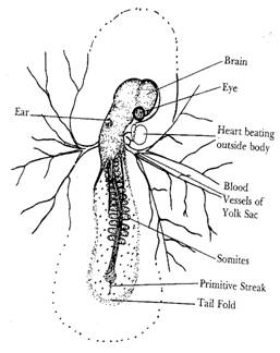
- DAY 2: Tissue development very visible. Appearance of blood vessels.
- DAY 3: Heart beats. Blood vessels very visible.
- DAY 4: Eye pigmented.
- DAY 5: Appearance of elbows and knees.
- DAY 6: Appearance of beak. Voluntary movements begin.
- DAY 7: Comb growth begins. Egg tooth begins to appear.
- DAY 8: Feather tracts seen. Upper and lower beak equal in length.
- DAY 9: Embryo starts to look bird-like. Mouth opening occurs.
- DAY 10: Egg tooth prominent. Toe nails visible.
- DAY 11: Cob serrated. Tail feathers apparent.
- DAY 12: Toes fully formed. First few visible feathers.
- DAY 13: Appearance of scales. Body covered lightly with feathers.
- DAY 14: Embryo turns head towards large end of egg.
- DAY 15: Gut is drawn into abdominal cavity.
- DAY 16: Feathers cover complete body. Albumen nearly gone.
- DAY 17: Amniotic fluid decreases. Head is between legs.
- DAY 18: Growth of embryo nearly complete. Yolk sac remains outside of embryo. Head is under right wing.
- DAY 19: Yolk sac draws into body cavity. Amniotic fluid gone. Embryo occupies most of space within egg (not in the air cell).
- DAY 20: Yolk sac drawn completely into body. Embryo becomes a chick (breathing air with its lungs). Internal and external pipping occurs.
The normal hatching position is:
- forepart of the body towards the large end of the egg;
- head under the right wing;
- legs up under the head.
Embryonic communication
With natural incubation, the chicks hatch over a relatively short period of time. This is despite the eggs being laid in the nest over a period of several days and the hen sitting on different eggs for different periods of time. This indicates that there is some system to synchronise the hatching process. It is now known that the different embryos communicate with each other by a series of clicking sounds, the rate of clicking being the important feature. Ensuring the eggs on the hatching trays are in contact with each other facilitates the synchronisation of hatching where the eggs are incubated in a modern machine. This helps to reduce the time between when the first and last chicks hatch.
References
- Anderson Brown, AF (1982) The Incubation Book, Saiga Publishing Company, Surrey, UK.
- Bradley, OC (1960) The Structure of the Fowl, Tom Grahame ed, Oliver and Boyd, Edinburgh, UK.
- North, MO and Bell, D (1990) Commercial Chicken Production Manual: 4th Edition, Van Nostrand Reinhold, New York, USA.
You May Also Like

The Poultry Hub Australia profoundly acknowledges and respects that its foundations, both people and facilities, are established on land rich in the history and traditions of the world’s oldest living culture. PHA values and respects Indigenous knowledge, understanding its importance in our shared history. We acknowledge the strength, resilience, and contributions of the Aboriginal community, we pay our tributes to the Aboriginal Elders – those who guided us in the past, those who lead us today, and those who will enlighten our paths in the future.