Digestive system
The Digestive System
The digestive system in the domestic fowl is very simple but efficient when compared to many other species, such as cattle. In the process of evolution, those avian species that developed simple but effective digestive systems were more able to fly and hence survive, as the simple digestive system would be lighter in weight. It is necessary that the diet provided to fowls be of high quality and easily digestible due to the simplicity in the structure and function of their digestive system. This is especially important if the birds are to attain the expected productive performance. This page describes the structure and function of the various parts of the digestive system of the fowl and discusses the digestion of poultry food into its constituent nutrients. The metabolism of carbohydrate, fat and protein is closely linked to the digestive system.
The digestive system consists of the alimentary canal along which the food passes after eating to where the residual wastes are eliminated from the body, together with the liver and the pancreas. The digestive system is responsible for the ingestion of food, its breakdown into its constituent nutrients and their absorption into the bloodstream, and the elimination of wastes from this process.
The liver produces bile and is associated with the metabolism of nutrients together with a number of other functions. The main function of the pancreas is the production of digestive enzymes and special compounds called hormones.
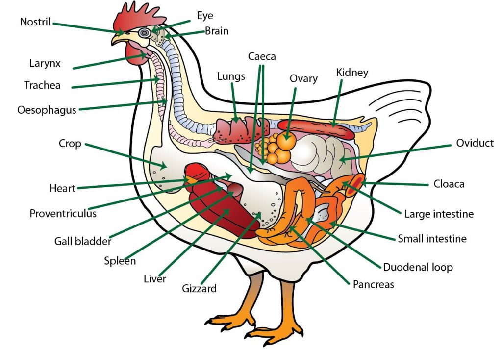
Anatomy of the chicken
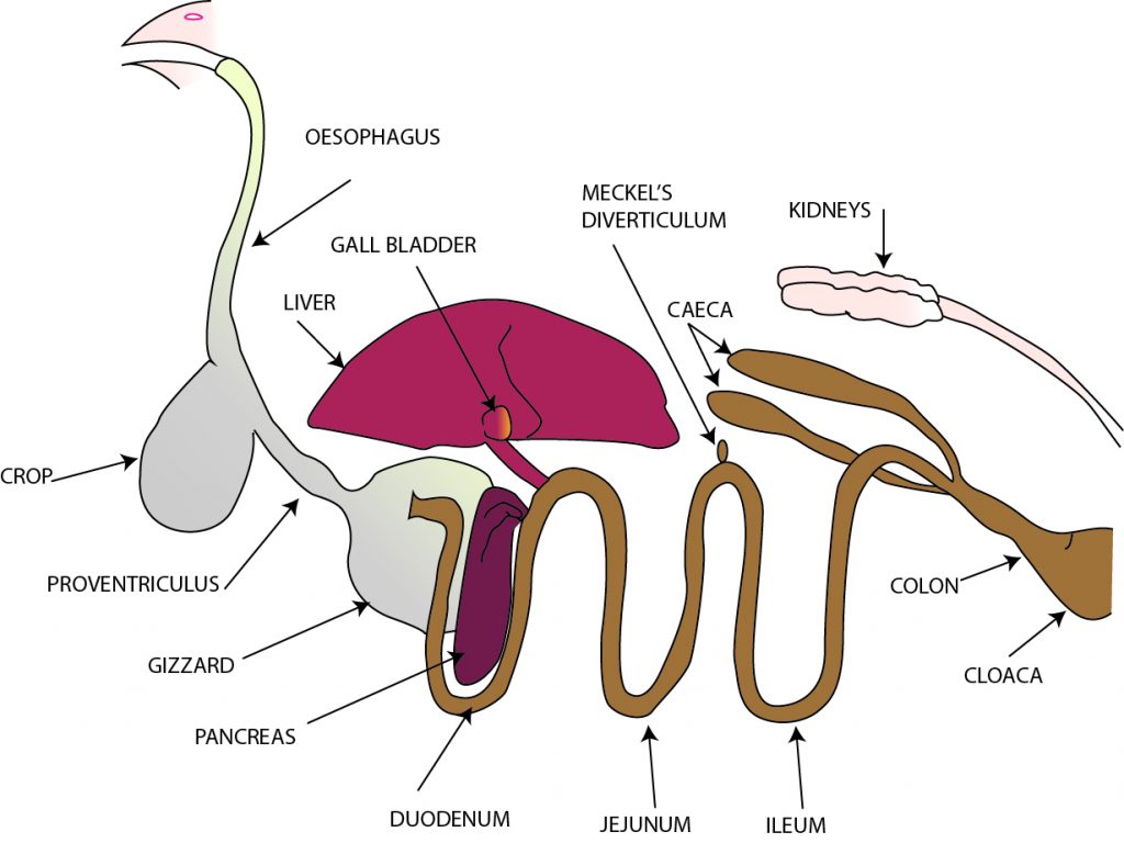
The alimentary canal
The alimentary canal is a long tube-like organ that starts at the beak and ends with the vent or cloaca in the abdominal region. Generally the alimentary canal has layers of muscle that run lengthwise and around it and is lined with mucous membranes. Glands that produce important digestive juices are found in different locations of the canal. The nutrients from the food, after digestion, are absorbed through the wall of the alimentary canal into the circulatory system for transport to the liver or other parts of the body. The waste remaining is eliminated from the body via the cloaca or vent.
Mouth structure
Fowls don’t have lips and cheeks, they instead have a beak which is an area of dense and horny skin lying over the mandible and incisive bones that serve as the bony foundation. There are no teeth. The so called egg tooth found on the end of the beak of newly hatched chickens is an aid to their escape from the egg at hatching and disappears after a day or two. The hard palate that forms the roof of the mouth, presents a long, narrow median (median – along the middle) slit that communicates with the nasal cavity. The hard palate has five transverse rows of backwardly pointing, hard, conical papillae. Numerous ducts of the salivary glands pierce the hard palate to release their secretions into the mouth cavity.
Salivary glands
A thick layer of stratified squamous epithelium covers the free surface. The salivary glands run the whole length of the hard palate, the groups of glands merging to form one mass of glandular tissue under the epithelium. Lymphoid tissue is found in most glands.
The salivary glands are:
- Maxillary – in the roof of the mouth
- Palatine – on either side of the nasal opening in the roof of the mouth
- Apheno-pteryoid glands – in the roof of the pharynx on each side of the common opening for the eustachian tubes (the eustachian tubes connect the middle ear to the mouth and their function is to equalise the air pressure on each side of the tympanic membrane in the ear)
- Anterior sub-mandible glands – in the angle formed by the union of the upper and lower beaks or mandibles
- Posterior sub-mandibular glands
- Lingual glands – in the tongue
- Crico-arytenoid glands – around the glottis
- A small gland in the angle of the mouth
Pharynx and tongue
The pharynx is continuous with, and follows, the mouth. The combined cavity of the mouth and the pharynx is often referred to as the oropharynx. The common opening for the two eustachian tubes is located in the middle of its dorsal wall (roof). The tongue is long and pointed and conforms to the shape of the beak in which it operates. The epithelium of the tongue is thick and horny, especially towards the tip. A transverse row of simple, large and horny papillae with their tips directed towards the rear of the mouth cavity are located on the posterior end. The hyoid bone provides the framework to support the tongue. The entoglossal bone extends longitudinally in the median plane. Small patches of lymphatic tissue are located throughout the corium. Mucous glands are located in the tongue with short ducts directed towards the rear. Some believe that there are taste buds located on the tongue, but this belief is not universally held. In any case, the sense of taste appears to be very weak if at all present.
The mouth has two major functions:
- To pick up the food particles.
- To direct the food into the oesophagus – as part of the bird’s eating behaviour.
Oesophagus, crop and proventriculus
The oesophagus is wide and is capable of being significantly stretched. It connects the mouth region to the crop in close association with the trachea. The crop is a large dilation of the oesophagus located just prior to where the oesophagus enters the thoracic cavity. The crop provides the capacity to hold food for some time before further digestion commences. This capacity enables the bird to take its food as “meals” at time intervals but permits continuous digestion. Inside the thoracic cavity, the oesophagus enters or becomes the proventriculus which is a very glandular part of the digestive tract (often called the glandular stomach).
The wall of the oesophagus is composed of four layers of tissue, the innermost being mucous membrane. The mucous membrane is an important barrier to the entry of microbes and the mucous it produces is a lubricant that aids the passage of the food along the alimentary canal. The structure below the crop is similar to that above except there is less lymphoid tissue below the crop. The crop structure is similar to that of the oesophagus except there are no glands present in fowls. Ducks and geese have glands in the crop mucous membranes. In pigeons the surface cells of the crop slough off during brooding to form pigeon’s milk which is used to feed the baby pigeons in the nest.
Proventriculus
The glandular stomach, or proventriculus, is relatively small and tubular. The wall is very thick and is composed of five layers:
- Outer serous membrane
- Muscle layer composed of three separate layers:
- Two thin longitudinal layers
- Thick circular layer
- Layer of areolar tissue containing blood and lymph vessels
- Thick layer composed mainly of glandular tissue
- Mucous membrane
The glands form the greater part of the thickness of the organ. Simple single glands group to form lobules each of which converges into a common cavity near the surface. The cavities converge to form a common duct that leads to the surface through the apex of a small papilla (see figure below). These glands produce a number of juices or enzymes that are used in the digestion or breaking down of food into its constituent nutrients. The mucous membrane is raised into folds and between these folds are numerous simple tubular glands that produce hydrochloric acid as well as lymphoid tissue.
Gizzard
The muscular stomach or gizzard is located immediately after the proventriculus, partly between the lobes and partly behind the left lobe of the liver. It has a flattened, rounded shape somewhat like a convex lens, with one side slightly larger than the other. Each surface is covered by a glistening layer of tendinous tissue which is thicker at the centre and becoming thinner towards the edges. Under this outer layer there are located very powerful masses of red muscle. The inner surface is lined with a creamy-coloured, thick, horny tissue raised in ridges. The gizzard almost always contains quantities of hard objects such as gravel or other grit that aids in the disintegration of food, which is the primary function of the gizzard.
The entrance from the proventriculus and the exit to the duodenum are close together and dorsal in location. The gizzard consists of a number of layers of tissues, some of which contain straight tubular glands. The innermost layer is a strong, flexible skin that is able to withstand the potentially damaging effects of the muscular action grinding the food often in the presence of stones or other insoluble material. The glands of the gizzard produce a liquid which is a keratinised material that passes to the surface of the horny lining where it hardens to replace tissue worn away by the grinding action of the organ.
The small intestine
The small intestine begins at the exit from the gizzard and ends at the junction of the small intestine, caeca and colon. It is relatively long and has a constant diameter. Of the three parts of the mammalian small intestine, the duodenum, jejunum and ileum, only the duodenum can be easily distinguished in the fowl. There is no clear demarcation between the jejunum and ileum and the small intestine appears as one long tube. Much of the digestion of the food and all of the absorption of the nutrients takes place in the small intestine and hence its structure is quite important. The structure is as follows:
- Serosa – a serous membrane on the outside of the intestine.
- A layer of longitudinal muscle – fibres run along the length of the intestine.
- A layer of circular muscle – three times as thick as the longitudinal muscle. Located between the two muscle layers are:
- Blood vessels
- Lymph vessels
- A network of nerve fibres
- An ill-defined sub-mucosa – the areolar of the oesophagus.
- Mucous membrane consisting of:
- A thick muscularis mucosae of longitudinal and circular muscle
- Corium – many glands, lymphoid tissue, muscle fibres and a variety of free cells
- Epithelium or surface
The small intestine has a number of very important functions:
- Produces a number of enzymes involved in the digestion process
- Site of much of the digestion of the food
- Site of much of the absorption of food
Villi
When a piece of the small intestine is immersed in water it takes on a very velvety appearance because of the presence of villi – long flattened, fingerlike projections that extend into the lumen (inside) of the intestine like flexible fingers. The villi are very actively involved in the absorption process. A single layer of columnar epithelium together with goblet cells covers the lining. The goblet cells secrete mucous. Permanent folds in the mucous membrane called the “valves of kerkring” are located at the proximal end (closest to the front) of the duodenum.
A lacteal (lymph vessels), capillaries, bundles of plain muscle fibres, nerves and other tissues and cells occupy the core of the villus. The villi have the function of providing a vastly increased surface area for the more efficient absorption of the nutrients. The efficiency of the absorption is influenced by the surface area available for the nutrients to move through i.e. the more villi the better the absorption. They also provide a means of concentrating the nutrients collection ability once they have moved through the intestine wall.
Duodenum
After the duodenum, the small intestine forms a coil and is suspended from the dorsal wall of the abdominal wall by a thin membrane called the mesentery. This membrane carries the blood vessels associated with the intestine. The duodenum starts at the gizzard and forms an elongated loop that is approximately 20 centimetres long. The pancreas lies between the arms of the loop and is attached to, and actually holds together, each arm of the duodenum.
Lymphoid tissue in the duodenum is very plentiful and is usually located in the corium. The lymphoid tissue collects the lymph and the lymph vessels transport fluid, other than blood, that is found in the spaces between cells and tissues until it passes into the blood system. Bile ducts from the gall bladder that are attached to the liver and two to three pancreatic ducts enter the small intestine by a common papilla at the caudal end (closest to the rear) of the duodenum. The pancreas is a very important organ in the process of digesting food and it is attached to each side of the duodenal loop and lies between the two arms.
Jejunum and the ileum
The jejunum and the ileum, together about 120 cm long, commence at the caudal end of the duodenum where the bile and the pancreatic duct papilla are located and terminates at the ileo-caecal-colic junction. This junction is where the small intestine, the two caeca and the colon all meet. This portion of the small intestine is similar in structure to the duodenum except that:
- It is suspended in the mesentery
- The villi are shorter
- There is less lymphoid tissue
Meckel’s Diverticulum is a constant feature about halfway along the small intestine and appears as a small projection on the outer surface of the small intestine. This projection is where the yolk sac was attached during the development of the embryo.
Large intestine
The large intestine is very short and does not differ to any extent from the calibre of the small intestine. It runs in nearly a straight line below the vertebrae and ends at the cloaca. Sometimes this section is referred to as the colon and the rectum (the rectum being the terminal section). The bursa of fabricius is located immediately above the cloaca of young birds but disappears when the birds have reached approximately one year old.
Caeca
The two caeca or blind pouches are about 16-18 centimetres long in the adult. They extend along the line of the small intestine towards the liver and are closely attached to the small intestine along their length by the mesentery. Each caecum has three main parts:
- A narrow base with thick walls arising at the ileo-colic-caecal junction
- Middle part with thin walls
- The wide blind apex with fairly thick walls
The structure of the caeca is as follows:
- Serous membrane
- Outer longitudinal muscle
- Circular muscle
- Inner longitudinal muscle forming the muscularis mucosae of the mucous membrane
Cloaca
The large intestine terminates in the front part of the cloaca. The cloaca is a tubular cavity opening to the exterior of the body and is common to the digestive and urogenital tract. The structure of the cloaca is very similar to that of the intestine except that the muscularis mucosa disappears near the vent. It divides into three chambers, each separated by a constriction not readily defined:
- The copradaeum – a continuation of the colon-rectum
- The urodaeum – middle part into which the ureters and genital ducts open
- The proctodaeum – opens to the exterior of the vent. Birds less than one year old have a dorsal opening leading into the blind, rounded sac – the bursa of fabricius
Liver
The liver is a bi-lobed organ that lies ventrally (below) and posterior (in rear of) to the heart and is closely associated with the proventriculus and the spleen. The right side lobe is larger. The liver is dark brown or chocolate in colour except for the first 10-14 days when it may be quite pale due to the absorption of lipids (fats) from the yolk as an embryo. It weighs approximately 50 grams in adult birds. The capsule, or glissosis, is the membrane that covers the liver and is thinner than that of mammals.
The gall bladder lies on the right lobe beneath the spleen. Two bile ducts emerge from the right lobe and one of these originates from the gall bladder and the second provides a direct connection from the liver to the small intestine. A system of ducts connects the right and left lobes.
There are a number of functions that are performed by the liver:
- Bile formation – consisting of bile, various pigments and bile salts. Bile is involved in the digestion of fats to fatty acids and glycerol
- The metabolism of:
- carbohydrate
- lipids
- protein
- Production and destruction of blood cells
- Synthesis of plasma proteins and fibrinogen (associated with blood clotting)
- Storage of glycogen, fat and fat-soluble vitamins e.g. vitamin A
- Detoxification of certain substances (detoxify – destroy the poisonous effect)
The liver cells have a high rate of destruction and a good regenerative capacity (re-growth ability). Notwithstanding this, in the normal animal, much of the organ is in reserve and can be removed or destroyed without causing undue stress.
Blood supply and drainage
There are two blood supply systems. One originates from the coelic artery for normal maintenance of the liver as an organ and the second, called the hepatic portal system, transports the nutrients from the small intestine after absorption to the liver. This latter system enters the liver via two veins (one for each lobe). The two blood supply systems join together inside the organ.
The liver is drained via the hepatic veins into the posterior vena cava (hepatic – relating to the liver; vena cava – one of the main veins that enters the heart). The liver has a network of sinusoids (empty holes in the tissues as in a sponge). The hepatic portal system, the capillaries of the arterial blood supply and the hepatic veins are in close association with each other in these sinusoid.
Bile
The liver consists of a series of tissue sheets that are two cells thick, with a sinusoid on either side of the sheet. Bile is made by the cells. The blood vessels, when they enter these sinusoids, become closely associated with them to provide for the easy transfer of material from one system to another. Minute canals called canaliculi that have the task of collecting and transporting the bile are associated with the cells in the tissue sheets. These canals eventually join together to form the bile ducts with one going directly to the intestine and one to the gall bladder before it connects to the small intestine.
Pancreas
This organ has three lobes that occupy the space between the two arms of the duodenal loop. Two or three ducts pass the secretions of this organ into the distal end of the duodenum via papillae common with the ducts from the gall bladder and the liver. The structure is similar to that of the pancreas of mammals and consists of special secreting tissue for pancreatic juice as well as other groups of cells called the “islets of Langerhans”. These are mainly associated with the production of hormones. In poultry, the cells of the islets of Langerhans are less defined than those in mammals. The functions of the pancreas are:
- Produce pancreatic juice which is a mixture of digestive enzymes.
- Produce the hormones insulin and glucagon that are involved in the metabolism of carbohydrate.
Interesting information about chicken digestion
The pattern of food intake and its passage through the digestive system are the main factors that influence secretory and hence digestive activity. Probably because of the high metabolic rate of the fowl, a more or less continuous supply of food is required by the digestive system. This is provided for by the crop that acts as a reservoir for the storage of food prior to its digestion and consequently permits the fowl to eat its food as periodic meals. There is quite wide variability between birds in relation to eating behaviour, even between those in the same flock. Some eat small amounts at short intervals while others eat larger amounts at wider intervals.
Food storage
The food is delivered into the crop for storage after the first few boli have passed into the proventriculus. The crop is quite distensible and will hold a large amount of undigested food that is then moved on as required by the proventriculus. This function of the crop is less important when there is a plentiful supply of food available. Due to the crop’s ability to hold a supply of food, when applying a food control (restriction) program, it is necessary to compensate by providing a long period of food deprivation to achieve the required degree of control. There is no relationship between the length of time of food deprivation and the amount of food consumed.
Food intake
While there is a wide variation between the eating habits of different birds in the flock, fowls do tend to eat meals on about 15-minute intervals through the daylight hours and, to some extent, during darkness. They tend to eat larger portions at first light and in the late evening.
Factors that affect food intake include:
- Age and live weight
- Environmental temperature
- Energy content of the food
- Food texture
- Level of other key nutrients
- Egg production
- Water quality and temperature
- The health status of the flock
Similar factors affect the rate of movement of the food through the digestive system with a meal of normal food taking approximately 4 hours to pass through in the case of young stock, 8 hours in the case of laying hens and 12 hours for broody hens. Intact, hard grains take longer to digest than the cracked grain and, quite often some whole grain will pass through unchanged.
Enzyme action
After ingestion, the food is mixed with saliva and mucous from the mouth and oesophagus and these secretions thoroughly moisten the food. The enzyme amylase, which is produced by the salivary and oesophageal glands and found in the saliva and mucous, can now commence to breakdown the complex carbohydrates. However, the amount of enzyme action at this stage is minimal and the first major enzyme activity takes place in the proventriculus and in the gizzard.
Hydrochloric acid, pepsin and gastrin
The secretions of the proventriculus, or glandular stomach as it is often called, include hydrochloric acid to lower the pH of the system and the food mixture, the enzyme pepsin that acts on protein, and the hormone gastrin that stimulates the production and release of gastric juice in the proventriculus and pancreatic juice from the pancreas.
Breaking the food particles
The gizzard is a very powerful organ which physically breaks the food particles into smaller sizes to make the work of the enzymes easier. At the same time, the enzymes previously released into the food with the saliva and by the proventriculus are thoroughly mixed into the food which improves their opportunity to carry out their work. This breaking and mixing function of the gizzard is enhanced by the presence of insoluble grit such as stones.
The food material enters the duodenum from the gizzard. Enzyme activity in this region is, in the main, a continuation of the breakdown of proteins started in the gizzard. Pancreatic juice and bile from the liver enters via ducts located at the distal end of the duodenum at about the junction of the duodenum and the jejunum if it were differentiated. However, because of back flow of pancreatic juice and bile towards the gizzard, the actions of these secretions start earlier in the digestive process than would be expected by their entry point to the small intestine. One effect is an increase in the pH of the intestinal contents of the latter half of the duodenum from strongly to weakly acid.
- In addition to enzymes, the pancreas produces insulin and sodium bicarbonate. The insulin is involved in the maintenance of blood sugar levels while the sodium bicarbonate, which is strongly alkaline, will increase the pH of the intestinal contents.
Reducing complex food compounds
The small intestine also produces enzymes that plays part in the digestive process of reducing the complex food compounds eaten to the simple compounds or building blocks that can be absorbed across the intestinal wall for transport to the organ or location where either they will be further processed, stored or used. Food materials that escape enzyme action along this tract are subjected to bacterial breakdown in the caeca which provides a system of at least partial recovery of some nutrients.
Waste (faeces)
The remainder of the material consists of waste and undigested food and are mixed with the urine in the cloaca and eliminated from the body as faeces. The appearance of the faeces varies considerably, but typically is a rounded, brown to grey mass topped with a cap of white uric acid from the kidneys. The contents of the caeca are also discharged periodically as discrete masses of brown, glutinous material.
- The average daily production of faeces from laying hens is between 100 and 150 grams. These fresh droppings are approximately 75% water and will air dry under favourable conditions to approximately 30% water.
Summary of digestion and metabolism
The utilisation of nutrients from the diet is a key element in the normal functioning of the animal. The avian digestive system is a simple system and consequently, the diet must be of good quality and consist of easily digested ingredients if the bird is to perform at the level required on the modern commercial poultry enterprise. Good working knowledge of the system and how it carries out its functions is necessary for the effective management of the poultry flock and, therefore, a study of the digestive system and the process of digestion and metabolism is an important facet in the study of poultry husbandry.
Further information
- Bradley, OC (1960) The Structure of the Fowl, Tom Grahame ed, Oliver and Boyd, Edinburgh, UK.
- Dingle, JG (1990) Module 3: Metabolism: Nutrient Procurement and Processing, Study Book: Poultry Husbandry 1, DEC, USQ, Toowoomba, Australia.
- Neisham, MC, Austic, RE and Card, LE (1979) Poultry Production, 12th Edition, Lea and Febiger, Philadelphia, USA.

The Poultry Hub Australia profoundly acknowledges and respects that its foundations, both people and facilities, are established on land rich in the history and traditions of the world’s oldest living culture. PHA values and respects Indigenous knowledge, understanding its importance in our shared history. We acknowledge the strength, resilience, and contributions of the Aboriginal community, we pay our tributes to the Aboriginal Elders – those who guided us in the past, those who lead us today, and those who will enlighten our paths in the future.