Circulatory system
The Circulatory System
The circulatory system is responsible for the transport of the various essential compounds and other factors around the body, as well as the removal of the metabolic wastes that accumulate in the tissues from body activities, to the appropriate places. The compounds and other factors transported around the body are blood, nutrients, medications and antibodies to fight infection, the residue of worn out cells and the wastes of metabolism. There are times when undesirable compounds and factors are found in the system as well, such as poisons or toxins and disease causing organisms.
The circulatory system consists of a number of organs and an associated transport system. These include the heart, the blood vessels, the spleen, the bone marrow, the blood and the lymph vessels. The blood and vascular system develop very early in the life of the embryo as the nutrition of the rapidly developing embryo is urgent and a transport system is required to transport the nutrients to where they are needed. Evidence of the system can be seen within about an hour and the system is clearly defined and operating within two days. By day three, it is possible to see the beat of the embryonic heart with the naked eye.
Fluids that act as a carrier of the various compounds and factors form part of the system. Each of these body fluids has its own function. Only circulatory fluids are discussed here. There are two types of body fluids:
- Circulatory fluids (these move around):
- Blood
- Lymph
- Static body fluids (remain stationary):
- Intracellular fluid or cell sap.
- Intercellular, interstitial or tissue fluid.
- Cerebrospinal fluid of the central nervous system.
- Synovial fluid in the joints of the skeleton.
- Aqueous and vitreous humor in the eye (fluid).
- Endolymph and perilymph in the ear (cochlea).
The circulatory system consists of a number of organs that produce the blood cells and the organs that pump it around the body (the heart), and the blood vessels such as the arteries, veins, capillaries and lymph vessels that carry it.
After the arteries leave the heart they divide to ever smaller and smaller arteries to provide the many branches required to service the many systems, organs, tissues and cells. When they reach the tissues they divide into capillaries that are very small and very thin walled (one cell thick). This allows the nutrients and factors being delivered to move out of the capillary into the tissues and the material to be collected from the cell to enter. When they leave the tissue the capillaries rejoin to form veins until, when near the heart, the blood is moving through either the vena cava or the pulmonary veins.
The lymphatic system is a special fluid collecting system that collects the fluid left behind in the tissues by the capillaries and transports it to a region of the heart where it enters the vena cava.
Heart
In broad terms the heart acts as the pump that pumps in two directions:
- To the lungs where the carbon dioxide in the blood is removed and the oxygen replaced
- To the rest of the body to deliver the nutrients and oxygen to the cells and to collect wastes and carbon dioxide
The blood leaves the heart via arteries called the aorta (to the body) and the pulmonary artery (to the lungs). The blood always enters the heart via the vena cava vein (from the body) and the pulmonary vein (from the lungs).
Location and shape
The heart is located in the thoracic cavity between the two lobes of the liver and mainly in front of that organ. It is relatively large and is enclosed in a thin membrane called the pericardium. The avian heart has two atria and two ventricles (four chambers), as is found in mammals. In general shape, it is typically conical with its apex, or pointed end, directed to the rear and slightly left of middle. A shallow groove, visible externally, indicates the division between the relatively small atria in the cranial end and the quite large ventricles making up the largest part of the organ in the caudal end.
Atria and ventricles and fibres
The walls of the atria are thin while those of the ventricles are quite thick. This is because of the atria only have to move the blood from the atria to the ventricle while the ventricles are responsible for pumping blood around the body. Microscopically, the muscle of the heart is similar to that of mammals as the fibres are nucleated, striated and form a syncytium (a mass of cytoplasm with numerous nuclei). Numerous fibres called Purkinje fibres are found in the heart under the endocardium and in the myocardium adjacent to the larger arteries.
The large veins draining the body (the two cranial vena cava and the caudal vena cava) enter the right atrium. The two pulmonary veins from the lungs join before entering the left atrium. The opening from the right atrium into the right ventricle is crescent-shaped. A strong muscular plate replaces the membranous tricuspid valve found in mammalian hearts. The opening between the left atrium and left ventricle is circular in shape with membranous flaps that correspond to the bicuspid valves of mammals. The septum (wall) between the two ventricles is thin with an even thinner area, called the fossa ovalis, located near the centre.
Ventricles
The external wall of the right ventricle is much thinner than that of the left. At its base is the opening for the pulmonary artery with three pocket-like semi-lunar valves. The openings of these pockets face away from the heart. The wall of the left ventricle is very thick except at the apex (pointed end). This is to provide the structural strength required for the heavy workload of that segment as it pumps blood around the body through the aorta. This leaves the heart from the left ventricle through an opening with semi-lunar valves similar that of the pulmonary artery. Tendinous cords called the chordae tendonae prevent too much movement of the valve segments from the force of the pumping heart (prevent the valves turning inside out).
Heart blood supply
The heart has its own blood supply via the two coronary arteries which divide from the aorta shortly after that artery leaves the left ventricle. The autonomous nervous system controls the pumping action of the heart.
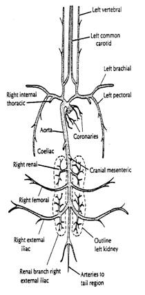
Arterial system
Arteries
The pulmonary system starts as with the single pulmonary artery that leaves the right ventricle and shortly after divides into the right and left branches. Each of these branches enter the lungs and divide to ultimately be involved in the gaseous exchange that takes place in the lungs. The aorta leaves the heart via the left ventricle and very shortly after divides to provide the two coronary arteries that supply blood to the heart itself. Shortly after that, the first of many divisions occur. Blood from the aorta is supplied to the head, the thoracic region including the wings, the abdominal region including the legs and tail.
New or oxygenated blood is bright red while old or deoxygenated blood is a very dark colour. In the pulmonary system, the arteries carry old blood while the veins carry new blood. In the main circulatory system, the arteries carry new blood and the veins carry old (arteries carry blood away from the heart and veins carry blood to the heart). As the arteries divide they become smaller and smaller until they form very small arteries which are often referred to as arterioles. When they enter the target organ they divide to become very thin walled capillaries and it is from these that the oxygen, nutrients, wastes and other factors either leave or enter the system.
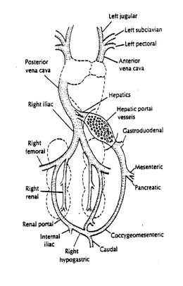
Veins
The capillaries in the various tissues and organs rejoin to form the veins that carry the blood back to the heart. Those draining the body, other than the lungs, enter the right atrium as three veins called:
- Caudal vena cava
- Right cranial vena cava
- Left cranial vena cava
The caudal vena cava drains the abdominal region while the two cranial vena cava drain the head via the juggler veins and the upper body including the wings and thoracic region excluding the lungs.
Kidney, small intestine and liver and lungs
The kidney system is very complex because of its function. The kidneys remove excess water and the wastes of metabolism from the blood stream. In addition, they need their own supply for the maintenance of their own tissues and cells. As in mammals, blood containing nutrients absorbed from the small intestine is carried to the liver by the portal vein. This is separate to the hepatic system that is responsible for the maintenance of the liver as an organ. The portal and hepatic systems join together in the liver to form the hepatic veins that enter the caudal vena cava near the heart. The capillaries in the lungs formed from the pulmonary arteries re-join to form the pulmonary veins that enter the heart via the left atrium for pumping to the rest of the body.
Blood
Venous system
Blood is the transport vehicle for the body. It is a very complex compound and consists of:
- Liquid intercellular substance or plasma
- Suspended formed elements
The formed elements include the red corpuscles or erythrocytes, the white corpuscles or leukocytes and the thrombocytes that correspond to the mammalian platelets. Blood also carries a great number and variety of other substances and factors. These include:
- Products from metabolism (wastes)
- Hormones
- Enzymes
- Antibodies
- Effete products of tissues and organs (effete = worn out)
- Many other organic and inorganic compounds
Erythrocytes
These are large, oval, flat, nucleated cells (mammalian erythrocytes are non-nucleated). Occasionally erytheroid structures without a nucleus are observed but these are normal avian erythrocytes from which the nucleus has been lost. The nucleus is centrally located and tends to be elongated in shape with a length of approximately 12 microns and a width of approximately 7 microns. The red colouring is caused by the presence of haemoglobin which is an iron compound that carries oxygen. The function of the erythrocytes is to transport the oxygen from the lungs to the tissues and the carbon dioxide from the tissues to the lungs. The erythrocytes are formed in the red bone marrow.
In the adult fowl there are 2.5 million erythrocytes in each millilitre of blood. The cell volume may be measured by taking a sample of the blood and centrifuging it at 3000 rpm for 15-20 minutes. The cells and the plasma separate and the volume of the cellular portion can be measured. This volume is called the haematocrit and, while it is mainly erythrocytes, it contains the leucocytes as well as a small quantity of plasma that has been trapped. The error as a result of the trapped plasma is approximately 5%.
Leucocytes
These are nucleated, amoeboid cells with a colourless cytoplasm. Some have fine granules in their cytoplasm, others carry coarse granules and others are non-granular. Because of the presence of granules the leucocytes are often called granulocytes. Most are phagocytic. The shape is therefore variable, but under normal conditions they are spherical. They are mono-nuclear although at times they appear to be multi-nuclear because of the shape of the nucleus as it is often multi-lobed.
The leucocytes are formed in the spleen, lymphoid tissue and in special cells in the bone marrow. The average count is approximately 30,000 per millilitre in adult birds and 10,000 per millilitre in day old chickens. Leucocytes may be classified by the character of their cytoplasm and nucleus. Protein granules in the cytoplasm may be acidic, basic or have neutral pH and this determines, to some extent, the type of stain they take up. Microscopic bodies such as cells and bacteria are usually stained in the laboratory to aid in their identification. Without staining, individually the different leucocytes are mainly colourless and it is very difficult to see their characteristics clearly even with a powerful microscope. Those identified in this way are:
- Acidic granules that stain with a basic (alkaline) stain and are called basophils.
- Basic (alkaline) granules that stain with an acidic stain and are called acidophils, oxyphils or eosinophils.
- Neutral granules may be stained with a neutral stain and are called neutrophils.
Using this system there are five main groups of leucocytes or granulocytes:
- Polymorphonuclear heterophilic granulocytes
- Polymorphonuclear eosinophilic granulocytes
- Polymorphonuclear basophilic granulocytes
- Lymphocytes
- Monocytes
Polymorphonuclear heterophilic granulocytes (heterophils)
These are known as heterophils. They are spherical cells with a diameter of 10-15 nanometres (nm). The nucleus is distinctly lobulated, with the lobes joined by thick strands of nucleoplasm (nucleus material). These cells play an important part in the body’s defence against bacterial invasion as they have a bactericidal function and ingest protein.
Polymorphonuclear eosinophilic granulocytes (eosinophils)
These are of similar size to the heterophils (10-15 nm), and the nucleus is frequently bi-lobed. The eosinophilic granules in the nucleus are round and are not so numerous as the granules in the heterophils. The function of the eosinophils is not definitely known but it is believed that they increase in number in cases of internal parasite infestations. It has been suggested also that they have a detoxifying function.
Polymorphonuclear basophilic granulocytes (basophils)
These are also about the same size as the heterophils and eosinophils. The nucleus is generally not lobulated but appears to be oval in shape. There tends to be fewer of these cells and their function is unknown, however it has been suggested that they are a stage in the degeneration of eosinophils.
Lymphocytes
These are the most numerous of all of the leukocytes. Their size varies significantly, the older cells being somewhat smaller than the newer cells because of the loss of cytoplasm from the older cells. The nucleus size is the same in all cells but may be kidney shaped in some cases. The mobility of these cells is greatly reduced and their phagocytic action is probably nil. Their function is thought to be that of regulating toxic materials. They also release enzymes that assist in the synthesis of nucleoplasm. In their degeneration they release special compounds called gamma globulins that have antibody properties.
Monocytes
Monocytes are the largest of all of the leukocytes. They are difficult to separate from the large lymphocytes because of a strong similarity in appearance. The nucleus may be round, oval or kidney shaped. They are very mobile and very phagocytic in their activity. Their function is to remove, by ingestion, bacteria, protozoan and tissue debris.
Thrombocytes
The thrombocytes are similar to the blood platelets of mammals, however, they are less involved with blood clotting. They are quite numerous (approximately 25000 per millilitre). They are the smallest of all of the blood cells and group together as small clumps. Thrombocytes are formed in the bone marrow in a way similar to the red blood cells.
Plasma
The plasma is the liquid or non-cellular portion of the blood. It contains a number of different compounds including:
- Glucose or blood sugar
- Non protein nitrogen substances
- Plasma proteins
- Plasma liquids
- Metallic elements
- Plasma enzymes
- Water
Blood sugar
The blood sugar of birds is in the form of D-Glucose as in mammals, but usually at higher, though variable, levels. Certain hormones such as insulin, glucagon, corticoids and glucocorticoid influence the actual level. The plane of nutrition is also an influence- Whether the bird is full fed or is starved (plane of nutrition) also influences blood sugar. The amount of sugar in the blood is approximately 260 mg/100 ml, although levels as low as 110 and as high as 350 mg/100 ml have been reported.
Non-protein nitrogen
After the removal of the plasma proteins, the remaining fluid contains a number of nitrogenous substances. The amount being in the approximate range of 15.5 mg/100 ml in the layer to 19.5 mg/100 ml in the immature bird. These include:
- Uric acid
- Urea
- Creatine
- Free ammonia
- Free amino acids
Uric acid and urea
Uric acid is the end product of protein metabolism and represents the bulk of the waste nitrogen eliminated by the birds. The level in the blood varies considerably and is influenced by the sex and reproductive state. As a result, non-layers have a higher level. Diet is another influence as birds on a high protein diet have a higher level of uric acid in their blood. Urea is another nitrogen containing compound and is eliminated by the kidney. Relatively small amounts may be found in the blood (2.2-2.5 mg/100 ml), and the level is influenced by age, sex and production.
Creatine, ammonia and free amino acids
Creatine is an amino acid present in animal tissues. It is equally distributed between the plasma and the cellular portion. It combines with phosphate to form phosphocreatine, an important compound in the anaerobic (without oxygen) phase of muscle contraction. The levels found range from 0.5-1.6 mg /100 ml with an average of approximately 1.0 mg/100 ml. Ammonia, or more specifically the ammonium ion, is found in such small quantities that further consideration is unwarranted. Free amino acids are found in very small quantities. About 22 different amino acids have been detected in the blood of fowls, but not all at the same time. The total amount has been estimated at approximately 8 mg/100 ml of blood.
Plasma proteins
Avian plasma contains a number of protein compounds which may be grouped into the following:
- Albumins – transport the fatty acids, calcium and other minerals, and hormones.
- Immunoglobulins – produced by lymphocytes as a result of antigenic demand.
- Transferrin – transports iron (Fe) across the intestine wall. Forms part of the secondary defence mechanism against bacteria and viruses by making iron unavailable to them.
- Ceruloplasmin – copper transport.
- Phosvitin – carries calcium, iron and phosphate to the yolk and calcium to the shell gland.
- Lipoproteins – carry fat-soluble vitamins, hormones and some minerals in the blood.
- Fibrinogen – is involved in blood clotting and is also present in the blood.
Lymph system
The body of the fowl is richly served with lymph vessels. Lymph is derived from the body fluid found within the interstitial space (the space between the important parts of the body). The lymphatic system has the function of draining the body systems of fluid that is left behind by the blood vessels, although the lymph fluid does ultimately return to the main circulatory system when the lymph vessel enters the vena cava near the heart. There are no lymph nodes in fowls. Lymph plexuses (an intertwining of the very small lymph vessels) are found instead of mammalian lymph nodes.
Spleen
The spleen is located immediately to the right of the junction of the glandular stomach (proventriculus) and the gizzard. The main function of the spleen is to filter out the unwanted particles from the blood. Another function is its involvement in the formation of lymphocytes. It is reddish brown in colour and generally round in shape. A thin, fibrous capsule with few muscle fibres surrounds it.
Summary of circulatory system and blood
The circulatory system is the main transport system of the body. It is the means by which nutrients, enzymes and other important needs for the proper functioning of body systems, organs, tissues and cells as well as body defence components are transported to where they are required. Waste products of tissue metabolism are also transported to where they will be eliminated or otherwise acted upon. Foreign material such as bacteria and viruses may be transported around the body by the system.
In order to carry out its function, the system consists of three main components – the pump (heart), the pathway (vessels) and the transport means (blood and lymph). Knowledge of how these components function and their relationship to the whole system is necessary to better understand the biology of the fowl as a whole.
Further information
- Bell, DJ (1971) Physiology and Biochemistry of the Domestic Fowl, Academic Press, London, UK.
- Dingle, JG (1991) Module 5: Internal Transport, Study Book – Poultry Husbandry 1; DEC; USQ, Toowoomba, Australia.
You May Also Like
The Poultry Hub Australia profoundly acknowledges and respects that its foundations, both people and facilities, are established on land rich in the history and traditions of the world’s oldest living culture. PHA values and respects Indigenous knowledge, understanding its importance in our shared history. We acknowledge the strength, resilience, and contributions of the Aboriginal community, we pay our tributes to the Aboriginal Elders – those who guided us in the past, those who lead us today, and those who will enlighten our paths in the future.