Reproductive system
The Reproductive System
The avian reproductive system is heterosexual and requires both a male and a female, each to contribute half of the genetic constitution of the offspring. The male contributes his half by way of the sperm produced by the testes and carried in the semen. The female contributes hers in the ovum carried by the egg yolk produced by the ovary. The ovum is often referred to as the blastodisc, blastoderm or germ disc. After release from the follicle on the ovary, the yolk moves into the oviduct where it is fertilised and has added to it the albumen, shell membranes and shell.
Male reproductive system
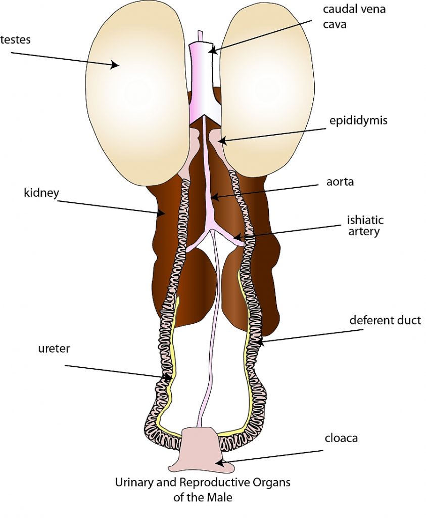
The urinary and reproductive organs of the male chicken
The male reproductive organs in the domestic fowl consist of two testes, each with a deferent duct that leads from the testes to the cloaca. Fowls do not have a penis such as is found in other animals. The testes are bean shaped bodies located against the backbone at the front of the kidney. Their size is not constant and they become larger when the birds are actively mating. The left testes is often larger than the right. On the inside of each is a small, flattened area that is believed to correspond to the epididymis of mammals. The deferent duct starts at this flattened area.
Deferent duct
The deferent duct transports the sperm from the testes where they are formed to the cloaca from which they enter the oviduct of the female when mating. The deferent duct enters a small pimple-like structure in the cloaca. This structure equates to the mammalian penis and is much larger in ducks to form a penis like organ. The deferent duct is quite narrow at first but widens as it approaches the cloaca.
Testes and sperm
In the testes very twisted tubes called seminiferous tubules are found. It is in these tubules that a special process of cell division called meiosis and transformation produces the sperm. The sperm carry half of the total chromosomes required to produce an embryo. The mother provides the other half. One cubic millimetre of the fluid called semen produced by the male contains on average 3-5 million sperm. Under a microscope the sperm of the fowl will be seen to have a long pointed head with a long tail. The testes also produce hormones called androgens that influence the development of what are called secondary sex characteristics such as comb growth and condition, male behaviour and mating.
Female reproductive system
The female reproductive system in the domestic fowl consists of the ovary and the accompanying oviduct. While the female embryo in chicken has two sets of reproductive organs, only one of these, the left survives and reaches maturity to produce eggs. The single surviving ovary is located in the laying hen just in front of the kidneys in the abdominal cavity and is firmly attached to the wall of the cavity. The ovary is well endowed with blood vessels to ensure there is no hindrance to the transport of nutrients to the developing yolk.
Ovary
The ovary consists of a mass of yellowish, rounded objects called follicles, each containing an ovum or yolk. There are many such follicles but only a small number in comparison, will ever reach maturity to produce an egg. When the hen is in lay the ovary will be active. The size of the follicles will vary from very small to those approaching the normal yolk size in the egg which can be up to 40 millimetres in diameter, and will contain a fully matured yolk ready for release into the oviduct.
It is possible to find five stages of development in the active ovary:
- Primary follicles – follicles that have not yet commenced to grow
- Growing follicles
- Mature follicles – follicles ready or nearly so for release
- Discharged follicles – where the yolk has just been released
- Atretic follicles – those from which the yolk has been released some time ago
Yolk
It takes approximately 10 days for a yolk to develop from the very small to the normal size found in eggs and during this time it is contained in the follicle. The follicle acts as a sack during this period of development supplying it with the nutrients required for its growth. When a mature follicle is examined an elongated area virtually free of blood vessels will be found on the distal surface of it. This area, called the stigma, is where the follicle normally splits to release the yolk into the oviduct. If, for some reason, the follicle splits at other than the stigma, the numerous blood vessels that rupture will result in free blood being found in the egg i.e. a blood spot will form.
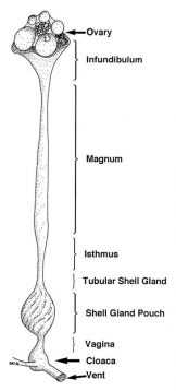
Ovary and oviduct
Oviduct
The function of the oviduct is to produce the albumen, shell membranes and the shell around the yolk to complete
the egg. It is a long tube well supplied with blood via numerous blood vessels. There are many glands found in its walls that produce the albumen, the shell membranes and the shell. In the non-layer, the oviduct is quite short and small in diameter. However, once the reproductive system becomes active, it grows to a length of 70-80 centimetres with a variable diameter depending on the function of the section being examined.
The oviduct consists of five distinct parts or sections, each having different functions:
- Infundibulum (or funnel): located adjacent to the ovary and with long segments enclosing the ovary, the infundibulum collects the yolk after its release from the follicle as a funnel and directs it into the oviduct. This section has very thin walls and is 6-9 centimetres long. Fertilisation of the ovum by the male sperm occurs here.
- Ampulla or magnum: at approximately 40 centimetres long it secretes more than 40% of the albumen.
- Isthmus: at about 12 centimetres in length, it secretes some albumen and the shell membranes.
- Uterus or shell gland: at approximately 12 centimetres in length it secretes about 40% of the albumen and the egg’s shell.
- Vagina: at approximately 12 centimetres in length, it secretes the egg’s outer cuticle and possibly the shell pigment.
Androgen, oestrogen and progesterone
In addition to the production of eggs, the female reproductive system also produces hormones that aid in the control of body functions. These include androgen, oestrogen and progesterone. Androgen causes comb growth and condition and has a function in the formation of albumen. Oestrogen causes the growth of the female plumage, mating and nesting behaviour, oviduct development together with the nutrient supply to the ovary/oviduct for egg formation. Progesterone, with androgen, is involved in the production of albumen and the carriage of the message to the pituitary gland to release luteinising hormone.
The female reproductive system remains dormant in the young chicken and growing pullet until she reaches the age when these organs start to prepare for the normal production of eggs. One of the first signs of her developing maturity is the change in the comb development. This organ starts to grow and to take on a vivid red hue as the hormones produced by the now awakening ovary start to have an effect.
The formation of the hen’s egg
The normal egg consists of the following major parts:
- Yolk carrying the ovum – produced by the ovary
- Albumen or white – produced mainly in the magnum
- Shell membranes – produced in the isthmus
- Shell – produced in the uterus or shell gland
The ovary and yolk formation
The ovary is attached to the abdominal cavity wall by the meso-ovarian ligament. It carries anything from 2,000 to 12,000 small ova in miniature follicles on its surface, plus hormone producing cells in its body. Not all of the ova found on the immature ovary develop and only approximately 200 to 350 reach maturity under normal modern commercial practice. Each yolk or ova takes about 10 days to grow and reach maturity when it is approximately 31% of the weight of the egg.
The composition of the yolk material is as follows:
| Component | % |
| Water | 48.0 |
| Protein | 17.5 |
| Fat | 32.5 |
| Carbohydrate | 1.0 |
| Other compounds | 1.0 |
The yolk is laid down in concentric rings of darker and lighter coloured material, the colour being produced by xanthophylls that are yellow/orange/red pigments occurring in many plants, plant products and other naturally occurring materials. The bulk of the yolk material provides a source of food for the developing embryo that originates by the fertilising of the germ disc or blastoderm usually located on the upper surface of the yolk of the broken out egg. It lies in the surface segment of the latebra which is a vase-shaped segment of different yolk with its base in the centre of the yolk, the lips on the surface and the stem joining the base to the lips.
Yolk development in the maturing pullet is initiated by follicle stimulating hormone (FSH) produced by the anterior lobe of the pituitary gland. The compounds in the yolk material are formed in the liver and, on the appropriate signal, are transported by the blood stream to the target follicle and into the yolk. The appropriate signal for this development comes from the hormones oestrogen, progesterone and testosterone which are produced by the ovary after receiving the signal of the FSH. These ovarian hormones also provide the stimulus for the formation of the development of the oviduct.
The yolk is contained in a very thin, transparent membrane called the vitelline membrane. As an egg becomes stale, the vitelline membrane becomes significantly weakened and often breaks to release the yolk contents when the stale egg is broken out. On ovulation the yolk is released and enters the oviduct where, as it passes along that organ, fertilisation occurs and the remaining parts of the egg are added around it. The yolk is located in a sack called the follicle, held on the ovary. The follicle, which although quite thin-walled, is extremely well supplied with blood vessels. These are necessary to carry the yolk constituting materials that have been formed in the liver.
Ovulation
The release of the yolk (the process of ovulation), is the major controlling factor influencing the subsequent steps in the formation and laying of the egg. As a consequence, factors that influence ovulation are of critical importance to the various aspects associated with egg production. The presence of a mature yolk in a follicle causes hormones from the ovary to stimulate the release of luteinising hormone (LH) by the pituitary gland. The presence of LH in the blood stream causes the follicle that contains the mature yolk to split along the stigma thus releasing it into the oviduct abdominal cavity adjacent to the oviduct.
Sexual maturity
Sexual maturity is reached when the hen lays the first egg in her life. Generally sexual maturity is genetically controlled, however, environmental factors play a very significant role. It will be in the age range of 18-24 weeks depending on fowl genotype, but it can be manipulated by controlled feeding practices, light intensity and day length management and other management practices.
Initiation of ovulation
The controlling mechanism setting the time of the day for the first ovulation is not fully understood. However, nervous and hormonal factors are important. Subsequent ovulations are, however, controlled largely by the time of the previous egg passing through the vent (being laid). Subsequent yolk release, if at all, occurs approximately 40-60 minutes after the previous egg has been laid.
Clutches
Eggs laid on successive days are called a clutch. Clutches are separated by days when no eggs are laid. Clutch size is an individual characteristic and may vary in a flock from 2 up to 100 eggs. However, the normal clutch size is significantly less than that and ranges from 3-8 eggs. The larger the clutch size the better will be the total production. Small clutch size indicates an inferior laying performance and is usually associated with long breaks in between.
Egg formation time
The time taken from ovulation until when the egg passes through the vent varies with individuals within the range of 23 to 26 hours. If the time is longer than 24 hours then the time of laying will be progressively later in the day for each successive egg in the clutch. When eggs are laid at a late hour, an ovulation is missed and the start of a new clutch will be earlier in the next laying day.
Ovulation time
Hens that produce long clutches release the yolk very shortly after first light (whether natural or artificial light). Successive ovulations occur very shortly after the laying of the previous egg. Those that produce short clutches usually release the yolk later in the day and often have longer periods between laying time and the next ovulation.
Laying pattern
When pullets first commence to lay, their hormonal and other controlling systems have not yet reached a state of balance. As a consequence, the first eggs are laid in a somewhat haphazard sequence. However, once these systems have reached a state of balance (usually after 7-10 days), egg production becomes more regular. Peak ovulation is reached 3-5 weeks after first egg. This will be held for a period and then will decline steadily thereafter until the bird moults or some other factor causes a cessation of production for a period.
Oviduct
The other components of the egg are the albumen, the shell membranes and the shell, and are produced by different segments of the oviduct. These segments are:
- Infundibulum
- Magnum or ampulla
- Isthmus
- Uterus or shell gland
- Vagina
- Cloaca
In the egg laying hen the oviduct is a tube like organ that consists of the previously named segments with one end lying adjacent to the ovary and the other entering the vent. It is approximately 70 centimetres long and is very glandular. The glands of the different segments produce the remaining different parts of the egg. Because of its function the oviduct is very well supplied with blood vessels.
Infundibulum
This segment is funnel-shaped and lies adjacent to the ovary. It is up to 9 centimetres long in the laying hen and has the function of searching for and engulfing the yolk that has just been released from the follicle into the adjacent ovarian pocket or body cavity. The yolk remains in the infundibulum for about 15 minutes and it is here that fertilisation takes place.
If the infundibulum malfunction and does not engulf the yolk, the yolk will remain in the ovarian pocket from where normally they will be absorbed within three days. If the number of such occurrences reaches a high level, the yolks will accumulate in the ovarian pocket faster than they can be absorbed. Such birds’ are called internal layers as the abdomen becomes distended and the hens adopt a very upright stance.
Magnum or ampulla
The magnum is the longest segment at up to 40 centimetres long. Its function is to add approximately 40% of the albumen to the developing egg that takes about three hours to move through. These percentages vary considerably depending on quite a few factors including the genetics of the hen, age of the bird, the egg’s age and/or storage conditions. However, in a good quality, freshly laid egg the above relationship mostly applies.
The chalazae are two twisted chords of albumen extending from the opposite sides of the yolk into the remaining albumen in the broken out egg. These two cords extend into the ends of the egg along the longitudinal axis and are parts of a very thin envelope of special albumen that surrounds the yolk and holds it in its position. The yolk has to remain centrally located for the survival of the embryo. The yolk turning or rotating as it passes along the oviduct causes the twisted effect of the chalazae.
While the bird produces only dense albumen, as the egg moves along the oviduct, water is added thus making liquid albumen. The rotation of the developing egg causes the albumen to separate into the inner liquid and the dense layers. The outer liquid layer is caused by the addition of more water when in the uterus. The dense layer contains significant amounts of mucin that binds it together in a jelly like form. As an egg stales, the amount of dense albumen decreases as it changes to the liquid form. The liquid form increases in volume and becomes even more fluid.
Albumen in a normal egg consists of four different layers as follows:
| Albumen layer | % |
| Chalazae and the chalaziferous layer | 2.7 |
| Liquid inner layer | 17.3 |
| Dense layer | 57.0 |
| Outer liquid layer | 23.0 |
Isthmus
The isthmus is approximately 12 centimetres long and has the functions of adding approximately 20% of the albumen and the shell membranes to the egg. There are two shell membranes:
- The inner shell membrane – laid down first
- The outer shell membrane – laid down last and about three times the thickness of the inner membrane
The isthmus takes approximately 75 minutes to carry out its tasks. While the egg is still in the oviduct the shell membranes appear as one over the total surface of the egg, so close, they are associated with each other. However, as the egg cools after it has been laid, the membranes separate, usually at the larger end to form the air cell. The air cell in the new laid egg is approximately 1.5 centimetres in diameter and approximately 0.5 centimetres deep.
As the egg ages, the interior contents lose water and the air cell increases in size. This change in size is an indicator of egg quality as related to the age of the egg and the holding conditions. The shell membranes consist of a fibrous protein material and act as a barrier to bacteria and fungi penetration into the egg. They also help reduce the rate of evaporation of water from the egg thus slowing the rate of deterioration of the egg. The isthmus also lays down the foundation for the shell by forming the first crystals of calcium carbonate on the outer shell membrane.
Uterus (shell gland)and eggshell quality
The uterus is a relatively short, bulbous gland up to 12 centimetres in length. The developing egg remains in the uterus for 18-20 hours while approximately 40% of the albumen and all of the shell is added. It is for this reason that the organ is often called the shell gland. Shell formation really begins by the deposition of small clusters of calcium carbonate crystals onto the outer shell membrane while in the isthmus. These are the initiation grains for the subsequent calcium carbonate deposition in the uterus. The number of these grains is genetically controlled and is related to the subsequent shell thickness as the more grains deposited in the isthmus, the thicker will be the final shell.
The shell of an egg is formed in two layers:
- Mammillary layer – a sponge like layer composed of soft calcite crystals (CaCO3). This layer is the inner layer.
- Palisade layer – formed of columns of hard calcite crystals; the longer the columns the stronger the shell. This layer is the outer layer of the egg.
The calcium for the eggshell comes from the diet, a special bone called medullary bone (found in the cavity of long bones)and the skeleton. The hen uses approximately 2.5 grams of calcium in the formation of one normal egg. She cannot absorb sufficient calcium from her diet each day (approximately 2.0 grams per day) to supply this need and hence, it becomes necessary for her to utilise skeletal calcium to make up the shortfall. This is particularly so at night when most of the shell is being formed but the hen in unlikely to be eating. In addition to the calcite, the shell also contains small quantities of sodium, potassium and magnesium.
The carbonate ions which go with the calcium to form the calcium carbonate of the egg’s shell, come from the blood and the shell gland. If anything should interrupt the supply of carbonate, thin-shelled eggs will result. This occurs in hot weather when hens pant to remove excess heat energy. The increased respiratory rate removes carbon dioxide from the blood thus reducing the carbonate ions available for eggshell formation.
Carbonic anhydrase is the enzyme which catalyses the conversion of carbon dioxide and water into carbonate ions. Zinc is the co-enzyme of carbonic anhydrase and any conditions resulting in Zn deficiency can lead to problems associated with egg shell formation.
There are many factors that influence eggshell quality:
- Length of time in lay: The longer the bird is in lay, the weaker the shells will become because of her inability to obtain enough daily calcium from her diet to supply all of her needs for one egg. As a consequence, better layers will deplete their skeleton calcium supply.
- Increased environmental temperature: This results in reduced food consumption (and calcium) and the reduction of carbonate ions because of panting.
- Egg laying time: Eggs laid early in the morning are more likely to have thinner shells than those laid by the same bird later in the day. This is because in the case of those eggs laid early the shells have been deposited during the hours of darkness when the bird does not eat, and therefore no dietary calcium for the shell formation.
- Stress: Stressed birds lay thinner shelled eggs.
- Body checked and misshapen eggs: Most of these defects are caused by the birds being startled shortly after the egg has entered the uterus and the first layers of calcium carbonate have been deposited. At this stage the shell is very fragile and weak and when startled the hen’s muscles contract (including those in the wall of the uterus) and thus crack the newly forming shell. These are covered by subsequent depositions of shell but the damage remains in the form of body checks and/or misshapen eggs.
- Disease: Certain diseases can cause weak shell and misshapen eggs.
- Drugs: Certain drugs influence eggshell formation and deposition.
The shell of an egg contains openings or pores. There are approximately 8,000 such pores in the shell of a normal hen’s egg. The function of these pores is to provide for the gaseous exchange during incubation and embryonic development. The developing embryo requires oxygen and gives off carbon dioxide. When the egg is first laid most of the pores are closed. However, as the egg ages more and more pores open up. The cuticle deposited on the outer shell is composed of organic material and water and blocks the pores. During the laying process the cuticle acts as a lubricant, but once laid, the egg’s surface soon dries and the residue, which is mainly protein, closes off most of the pores as a barrier to the invasion of bacteria and fungi.
Vagina
The vagina is about 12 centimetres in length. While not known for sure, it may have the function of adding pigment to the outer shell to provide the egg with its colour.
Cloaca
The egg is held in the cloaca immediately prior to being laid. It may be in the cloaca for several hours, but usually is held there for a much shorter time. Although the egg usually enters this organ small end first, it usually rotates there to be laid by the large end first. However, if the bird should be startled at this time the egg may be forcibly expelled small end first.
Summary of the reproductive system
A bird’s reproductive system permits early separation of the hen from her offspring, which permits the hen to fly and reproduce at the same time. The formation of an egg is a very complex activity during which much can go wrong. The quality of the final product, the egg as it is laid, is influenced by both genetic and management factors. A working knowledge of the fowl’s reproductive system and the formation of the egg assists the farmer to maximise egg production and quality.
Further information
- Bradley, OC (1960) The Structure of the Fowl, 4th Edition, Tom Grahame ed, Oliver and Boyd, London, UK.
- North, Mack O and Bell, D (1990) Commercial Chicken Production Manual, 4th Edition, AVI Publishing Company, USA.
- Parkhurst, CR and Mountney, GJ (1988) Poultry Meat and Egg Production, Von Nostrand Reinbold Company, Melbourne, Australia.
You May Also Like

The Poultry Hub Australia profoundly acknowledges and respects that its foundations, both people and facilities, are established on land rich in the history and traditions of the world’s oldest living culture. PHA values and respects Indigenous knowledge, understanding its importance in our shared history. We acknowledge the strength, resilience, and contributions of the Aboriginal community, we pay our tributes to the Aboriginal Elders – those who guided us in the past, those who lead us today, and those who will enlighten our paths in the future.