Muscular system
The Muscular System
The muscular system provides the mechanical activity for the animal in the form of mobility of the different parts of the skeleton or its appendages, the movement of materials along tubular organs such as the alimentary canal, air passages and blood vessels, and the pumping of the blood through the circulatory system by the heart.
Muscles are structured from special muscle cells in the form of fibres that have the ability to contract or shorten. When they relax the muscle lengthens.
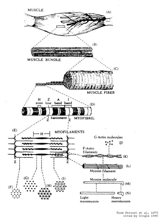
Components of a muscle
Muscle types
There are three types of muscle found in the animal body. These are:
- Involuntary muscles found in the walls of the alimentary canal, blood vessels, air passages and other tubular structures. These muscles are beyond the control of the will and are called involuntary muscles. The fibres of these muscles do not carry transverse striation or stripes and are therefore said to be ‘unstriped’ or ‘unstriated’.
- Cardiac muscle of the heart. This too is involuntary muscle but is striated and is structured differently to other muscle. It is nucleated, contains many Purkinje Fibres and forms a syncytium with many nuclei but no differentiation of the protoplasm into cells.
- The striated or striped, voluntary muscles of the body that move the various parts of the skeleton or appendages. These consist of very minute thread-like muscle fibres in bundles enclosed by sheaths of fibrous tissue.
Structure of a skeletal muscle
The typical voluntary muscle consists of bundles of long muscle fibres. Each fibre is made of long strands called myofibrils that in turn consist of segments called myofilaments. The myofilaments consist of myosin filaments and actin filaments arranged so that the myosin filament is a core surrounded by the actin filaments that are not continuous but form a cap-like structure at each end. Skeletal muscles are attached to the bones by very strong, fibrous bands or cords called tendons.
Muscle contraction
Muscle contraction occurs as a result of a stimulus usually originating from the nervous system either on a voluntary or involuntary basis. When muscle contraction occurs the myofibril segments shorten as a result of the actin filament caps sliding along the myosin filaments. Bands across the myofibril produce the striped effect of skeletal muscle. The muscular system provides the power for the animal and it is here that much of the energy contained in the diet is utilised for normal involuntary and voluntary activity.
Types of skeletal muscle fibres
There are white and red types of skeletal muscle fibres found in birds and all muscles have some white fibres and some red fibres. However, the proportion varies and some muscles are predominantly white and others predominantly red or dark. White fibres lack a compound called myoglobin, but store more glycogen and have a fast contraction of short duration. They have little staying power. The breast muscles of fowls, the muscles of flight, are predominantly white fibres and fowls have very poor flying ability. They fly very short distances with a very rapid wing movement. Red fibres have myoglobin and other cellular structures for continuous production of energy for contraction. These fibres have a slow contraction of long duration. The flight muscles of flying birds consist mainly of red fibres.
Rigor mortis
When an animal dies the characteristic energy reactions become irreversible and the muscles contract and do not relax for several hours. The stiffening effect of muscle contraction is called rigor mortis. This condition develops at a faster rate in warmer temperatures or if the animal is fatigued. Relaxation of the muscles usually occurs 10-12 hours after the onset of rigor due to subsequent changes in the body.
Further information
- Dingle, JG (1991) Animal Anatomy and Physiology 11(P), DEC, USQ, Toowoomba, Australia.
You May Also Like
The Poultry Hub Australia profoundly acknowledges and respects that its foundations, both people and facilities, are established on land rich in the history and traditions of the world’s oldest living culture. PHA values and respects Indigenous knowledge, understanding its importance in our shared history. We acknowledge the strength, resilience, and contributions of the Aboriginal community, we pay our tributes to the Aboriginal Elders – those who guided us in the past, those who lead us today, and those who will enlighten our paths in the future.