Excretory system
The Excretory System
The excretory system in the domestic fowl consists of the two kidneys, each with a ureter that carries the urine produced by the kidneys to the cloaca where it leaves the body. When the kidneys are diseased or damaged and unable to carry out their functions efficiently, the animal becomes debilitated and death often occurs quickly. Its functions in the domestic fowl are to:
- maintain the electrolyte balance
- maintain the water balance
- eliminate metabolic wastes, particularly nitrogen products of metabolism (except carbon dioxide)
The Kidneys
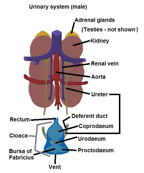
Urinary system - male
The two reddish-brown kidneys of the domestic fowl, each generally with three lobes, are found immediately behind the lungs on each side of the vertebral column and closely associated with it. A relatively straight, narrow tube called the ureter, leaves the medial border of each kidney and opens into the cloaca adjacent to the deferent duct of the male or the oviduct of the female. The ureter connects to many funnel shaped structures from each lobe of the kidney.
The kidney, on close inspection, consists of the renal cortex and the renal medulla. A microscopic examination of a section of kidney will show that it consists of a large number of renal tubules, or nephrons, each divided into cortical and medullary parts. Birds have two types of nephrons. A small number of ‘mammalian-type’ nephrons with a Loop of Henle (used to help concentrate the urine) found in the renal medulla and a much larger number of ‘reptilian-type’ nephrons without the Loop which are located in the renal cortex. The renal tubules extract the constituents of the urine from the blood that flows through the kidney.
A renal tubule or nephron is composed of the:
- renal corpuscle which consists of a close network of blood capillaries almost enclosed in a capsule called Bowman’s Capsule. It is here that the necessary very close association with the blood occurs.
- proximal convoluted (spiral-like) segment leading from Bowman’s Capsule
- the loop of straight tubule called the Loop of Henle (only in the mammalian-type nephrons).
- distal convoluted segment.
- collecting tubule that directs the urine into the ureter for elimination from the body.
Urine
Like almost all birds, the domestic fowl does not have a bladder as is found in most mammals and amphibians. The urine leaves the ureters and enters the cloaca where it is moved by reverse peristalsis into the large intestine, which permits excess water to be re-absorbed before elimination. This re-absorbed water is available for use by the bird and, to some extent, offsets the limited ability of birds to concentrate their urine as efficiently as mammals. The urine is in a thick pasty form with a very low water content but high in uric acid from nitrogen metabolism. It is usually passed as a paste and is deposited as a whitish or cream cap on some faecal stools.
When the kidneys are not functioning as efficiently as normal, or sometimes when a very high protein diet is provided, there will be large quantities of uric acid in the blood and the system may be unable to cope. The kidney tubules are likely to swell with accumulated urate deposits and when this happens, the white lines are clearly visible on the surface of the kidneys. The accumulation may lead to damage of the kidney cells which leads to nephritis. The high concentration of uric acid in the blood may result in filtration through the capillary walls which leads to visceral gout, which is when a whitish deposit is found on the surface of many visceral organs. Infectious bronchitis may produce these effects.
Further information
- Bradley, OC (1960) The Structure of the Fowl, Tom Grahame ed, Oliver and Boyd, Edinburgh, UK.
- Dingle, JG (1991) Animal anatomy and Physiology 11(P); Study Book, DEC, USQ, Toowoomba, Australia.
You May Also Like

The Poultry Hub Australia profoundly acknowledges and respects that its foundations, both people and facilities, are established on land rich in the history and traditions of the world’s oldest living culture. PHA values and respects Indigenous knowledge, understanding its importance in our shared history. We acknowledge the strength, resilience, and contributions of the Aboriginal community, we pay our tributes to the Aboriginal Elders – those who guided us in the past, those who lead us today, and those who will enlighten our paths in the future.